Pea Disease Diagnostic Series (PP1790, May 2016)
Roots and Wilts
Fusarium root rot
Fusarium avenaceum, F. solani f. sp. pisi and other species
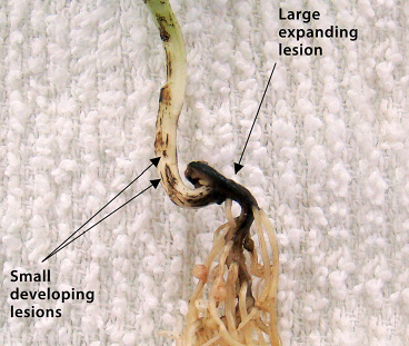
FIGURE 1 – Discrete lesions expanding from the point of seed attachment and coalescing into larger lesions
Photo: L. Porter, USDA-ARS Prosser, WA
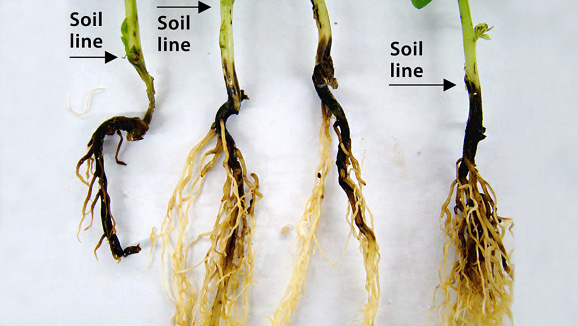
FIGURE 2 – Advanced lesions affecting large areas of roots and hypocotyls
Photo: L. Porter, USDA-ARS Prosser, WA
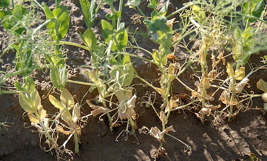
FIGURE 3 – Infected plants yellowing from the base upward
Photo: L. Porter, USDA-ARS Prosser, WA
AUTHORS: Julie S. Pasche, Lyndon Porter and Kimberly Zitnick-Anderson
SYMPTOMS
• Red to brown-black below-ground lesions
• Lateral root reduction and complete destruction in severe infections
• Below-ground red discolored vascular tissue is possible
• Above-ground stunting, yellowing and necrosis
FACTORS FAVORING DEVELOPMENT
• Temperatures from 73 to 83 F and wet soils
• Soil compaction and plant stress
• Contaminated seed or plant debris
IMPORTANT FACTS
• Alternative hosts include dry beans, soybean, chickpea and lentil
• Often seen in a complex with other root rots
• Above-ground symptoms often not seen until flowering
• Can be confused with other root rots and abiotic stress (water damage, etc.)
Aphanomyces root rot
Aphanomyces euteiches
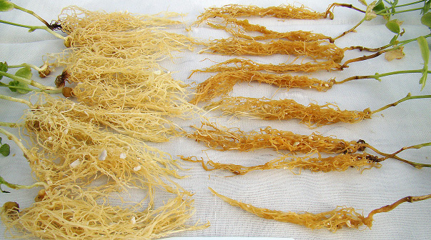
FIGURE 1 – Caramel-brown infected roots (R) and healthy roots (L)
Photo: L. Porter, USDA-ARS Prosser, WA
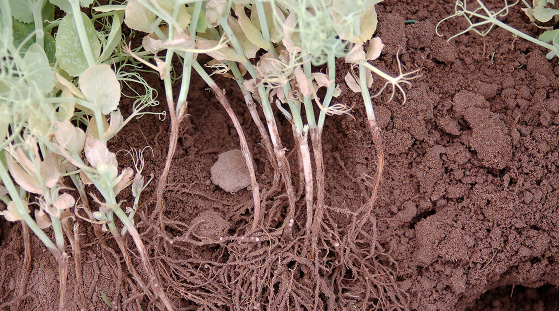
FIGURE 2 – Infected roots and yellowing lower leaves
Photo: L. Porter, USDA-ARS Prosser, WA
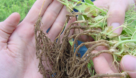
FIGURE 3 – Outer root tissue sloughing off and exposing inner vascular tissue
Photo: L. Porter, USDA-ARS Prosser, WA
AUTHOR: Lyndon Porter
SYMPTOMS
• Caramel-brown root and below-ground stem
• Outer root and below-ground stem tissue will slough off, exposing the vascular tissue
• Lower leaves turn yellow; the plant may be stunted, wilt and/or die prematurely
FACTORS FAVORING DEVELOPMENT
• Cool and wet spring conditions
• Low-lying areas
• Short rotations with peas or lentils
IMPORTANT FACTS
• Thick-walled spores can survive in soil for 20 years or more
• Lentils are a host, but chickpeas and faba beans are not
• Crop rotations of six or more years with nonhost can help reduce disease
• Can be confused with other root rots and abiotic stress (water damage, etc.)
Pythium seed and seedling rot
Pythium ultimum and other Pythium species
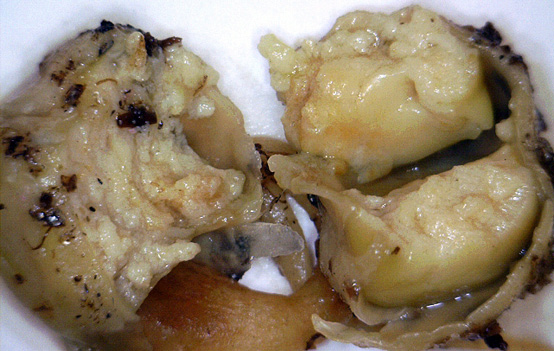
FIGURE 1 – Light brown internal seed rot
Photo: L. Porter, USDA-ARS Prosser, WA
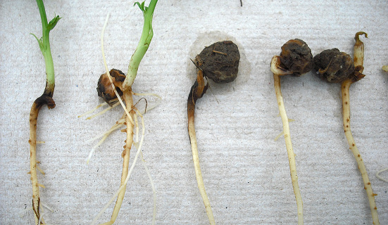
FIGURE 2 – Rotted seed coated with soil
Photo: L. Porter, USDA-ARS Prosser, WA
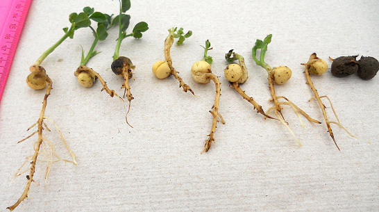
FIGURE 3 – Emerged plants with reduced vigor
Photo: L. Porter, USDA-ARS Prosser, WA
Rhizoctonia seed, seedling and root rot
Rhizoctonia solani AG 2-1, 4, 5 and 8
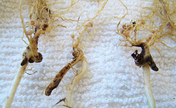
FIGURE 1 – Sunken brown lesions on below-ground stem tissue
Photo: L. Porter, USDA-ARS Prosser, WA
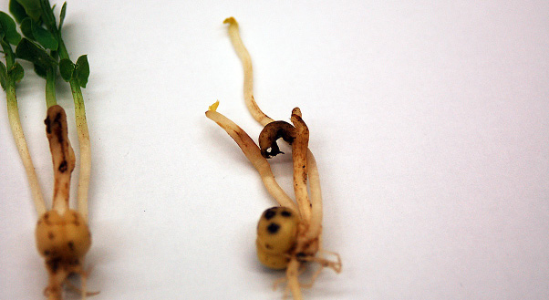
FIGURE 2 – Browning of the roots and pinching-off of root tips
Photo: L. Porter, USDA-ARS Prosser, WA
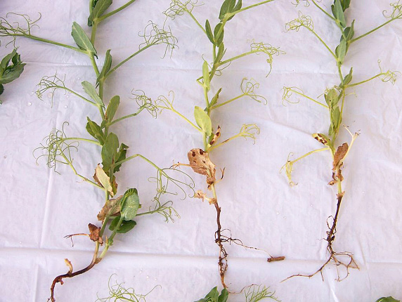
FIGURE 3 – Peas infected with Rhizoctonia
Photo: K. Chang, Alberta Agriculture and Forestry
AUTHORS: Timothy Paulitz, Dipak Sharma-Poudyal, Lyndon Porter, Weidong Chen and Lindsey du Toit
SYMPTOMS
• Seeds may rot in soil, resulting in poor emergence
• Seedlings have reddish-brown, sunken lesions on roots and base of stem
• Pinching-off of tips of the main tap root and secondary roots
• Plants become stunted and yellow
• Wet, cool soils
• Seed with poor germination
IMPORTANT FACTS
• Pathogen can survive in soil and plant debris
• Rotation is largely ineffective and resistant cultivars are not available
• Fungicide seed treatments are recommended
• Can be confused with other root rots, water damage
Fusarium wilt
Fusarium oxysporum f. sp. pisi
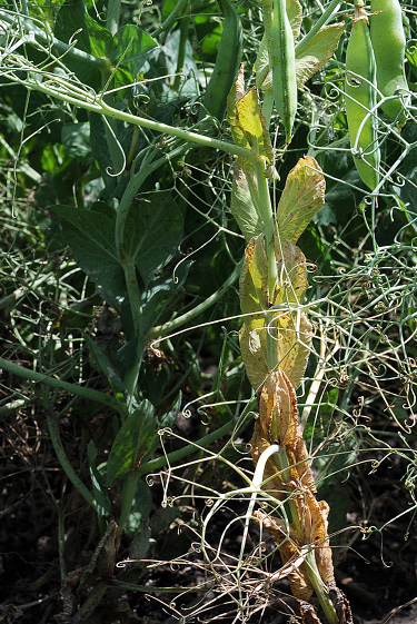
FIGURE 1 – Yellowing and curling of leaves
Photo: S. Markell, NDSU
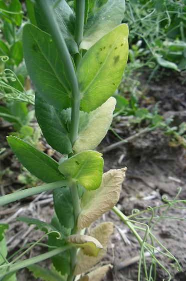
FIGURE 2 – Curling and yellowing of lower leaves on one side of the plant only
Photo: S. Guy, Washington St. U.
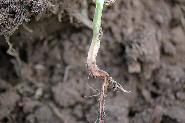
FIGURE 3 – Orange-red vascular discoloration extending into the stem
Photo: L. Porter, USDA-ARS Prosser, WA
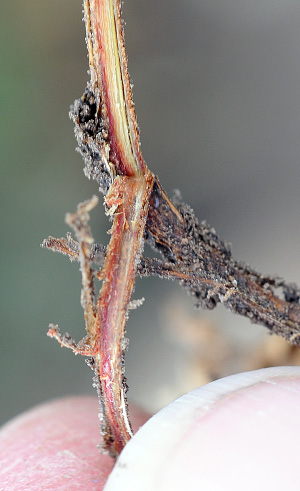
FIGURE 4 – Severe vascular discoloration
Photo: S. Markell, NDSU
AUTHOR: Stephen Guy
SYMPTOMS
• Leaves curl and yellow progressively from the base of the plant upward, sometimes more severe on one side of the plant
• Root vascular tissue is shades of yellow, orange or red, extending into the base of stem
• Field distribution is scattered plants or concentrated patches
• Plants may wilt
• Previous history of disease in the field
• Frequent cropping of susceptible varieties
• Late planting
IMPORTANT FACTS
• Can survive in soil for 10 years or more
• The fungus penetrates root tips and blocks vascular tissue
• Pathogen has more than one race and resistant varieties may not be effective against all races
• Can be confused with Aphanomyces and Fusarium root rots and abiotic stress
Spots and Lesions
Ascochyta blight
Ascochyta pisi, A. pinodes,
Phoma medicaginis var. pinodella
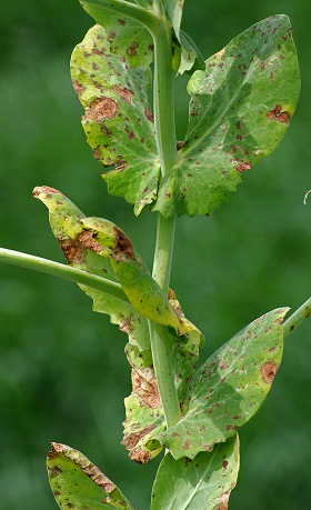
FIGURE 1 – Oval lesions with concentric rings
Photo: M. Wunsch, NDSU
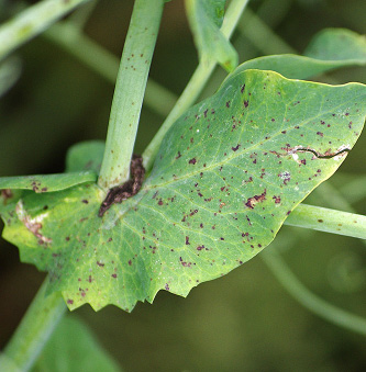
FIGURE 2 – Irregular flecks on leaf, extending to petioles and stems
Photo: M. Wunsch, NDSU
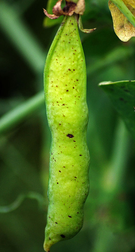
FIGURE 3 – Small, irregular pod lesions
Photo: M. Wunsch, NDSU
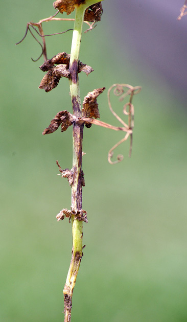
FIGURE 4 – Stem lesions
Photo: M. Wunsch, NDSU
SYMPTOMS
• Leaf lesions are dark, irregular flecks and/or circular to oval lesions, with a concentric ring pattern
• Purplish stem lesions develop at nodes, elongate and may girdle stem
• Pod lesions are small, irregular to circular and brown to purplish black
• Seed may be discolored
FACTORS FAVORING DEVELOPMENT
• Cool, wet weather
• Short rotational intervals between pea crops
IMPORTANT FACTS
• Primarily residue-borne but can be seedborne
• Crop rotation reduces but does not eliminate pathogen inoculum
• The host range of the causal pathogens is limited to field peas
• Can be confused with bacterial blight or Septoria blight
Bacterial blight and brown spot
Pseudomonas syringae pv. pisi and P. syringae pv. syringa
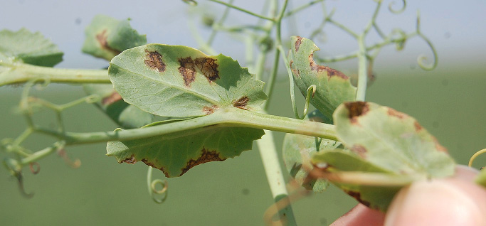
FIGURE 1 – Angular leaf lesions delimited by veins
Photo: R. Harveson, Univ. of Nebraska
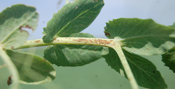
FIGURE 2 – Watery stem lesions forming in linear patterns as disease progresses
Photo: R. Harveson, Univ. of Nebraska
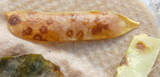
FIGURE 3 – Bacterial ooze emerging from pod lesions
Photo: R. Harveson, Univ. of Nebraska
AUTHOR: Robert M. Harveson
SYMPTOMS
• Symptoms occur on all above-ground plant parts
• Lesions initially are water-soaked and later turn necrotic
• Lesions are vein-delimited, angular in shape and translucent
• Bacterial ooze may be seen under conditions of high humidity
FACTORS FAVORING DEVELOPMENT
• Warm temperatures
• High humidity or leaf moisture
IMPORTANT FACTS
• Pathogens are seedborne
• Spread can occur with any type of mechanical contact on wet leaves or by splashing water
• Planting clean seed and use of disease resistant cultivars are the most effective management tools
• Can be confused with fungal leaf spots
Powdery mildew
Erysiphe pisi and E. trifolii
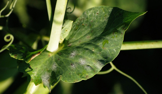
FIGURE 1 – Small tufts of fungal growth
Photo: M. Wunsch, NDSU
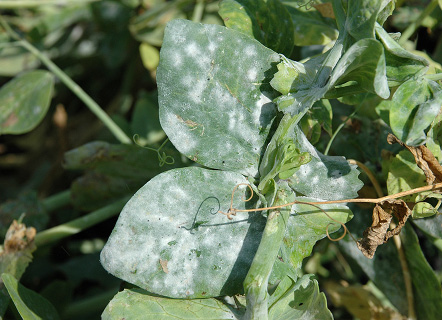
FIGURE 2 – Progression of fungal growth
Photo: S. Markell, NDSU
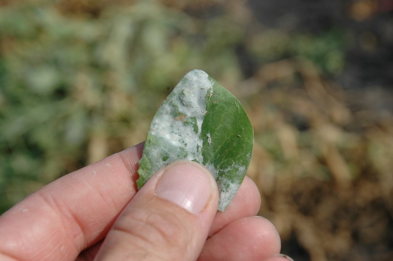
FIGURE 3 – Fungal growth rubbed off right side of leaf
Photo: S. Markell, NDSU
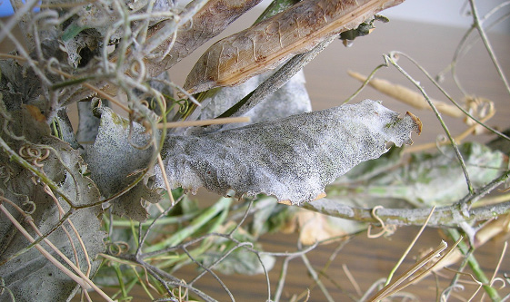
FIGURE 4 – Sever infection late in the season; note black fungal structures
Photo: R. Attanayake, Washington St. U.
AUTHORS: Renuka N. Attanayake, Weidong Chen and Michael Wunsch
SYMPTOMS
• White powdery tufts of fungal growth
• New fungal growth can be rubbed off easily
• Fungal growth will expand and may cause plant tissue to become chlorotic
• Late in the season, black fungal structures may appear
• Infection on pods can cause a gray-brown discoloration of the seeds
FACTORS FAVORING DEVELOPMENT
• Temperatures of 59 to 77 F are optimal
• Heavy dew or fog
• Late planting
IMPORTANT FACTS
• Pathogen can be soil-borne, seed-borne and wind-dispersed
• Management tools include resistant cultivars, crop rotation and foliar fungicides
• Most prevalent late in the season
Rust
Uromyces viciae-fabae
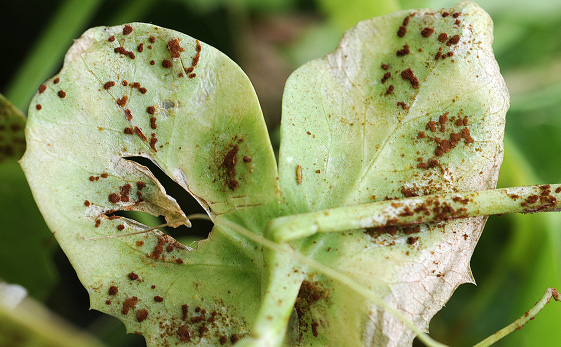
FIGURE 1 – Pustules filled with dusty brown spores on leaf
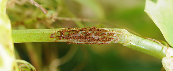
FIGURE 2 – Pustules lacerating branch
Photo: S. Markell, NDSU
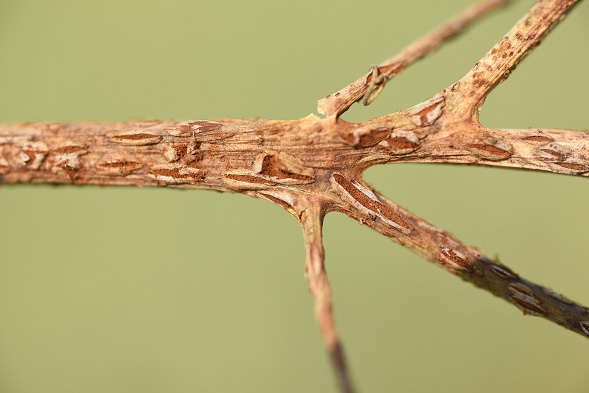
FIGURE 3 – Severe infection causing premature senesce and plant death
Photo: S. Markell, NDSU
SYMPTOMS
• Affects all above-ground plant parts
• Pustules erupt from tissue, causing holes and large lacerations
• Pustules are filled with dusty cinnamon-brown spore that easily rub off
• Severe infection causes yellowing, premature senesce and yield loss
FACTORS FAVORING DEVELOPMENT
• Heavy dew or fog
IMPORTANT FACTS
• Disease observed annually in northern Great Plains but rarely widespread
• Epidemics can progress quickly once disease is established
• Foliar fungicides can help manage disease
• Also can infect lentils and garden peas
Septoria blight
Septoria pisi
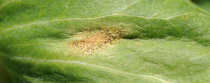
FIGURE 1 – Young leaf lesion with black fungal structures (pycnidia)
Photo: S. Markell, NDSU
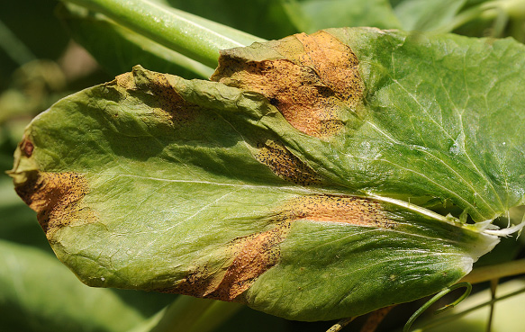
FIGURE 2 – Oblong lesions with pycnidia
Photo: S. Markell, NDSU
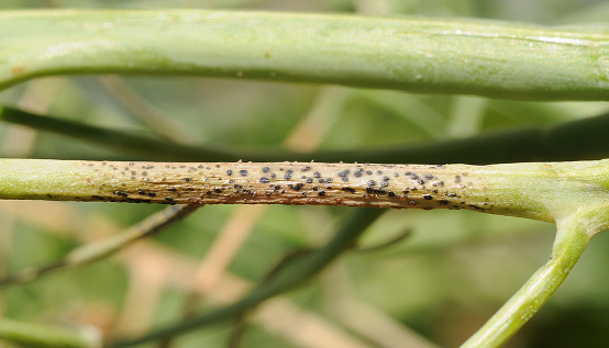
FIGURE 3 – Necrotic lesion with pycnidia on branch
Photo: S. Markell, NDSU
AUTHORS: Mary Burrows and Sam Markell
SYMPTOMS
• Symptoms occur on all plant parts
• Necrotic lesions with small black fungal structures (pycnidia)
• Often occur late in the season
FACTORS FAVORING DEVELOPMENT
• Warm temperatures (70 to 80 F)
• High humidity or heavy dews
IMPORTANT FACTS
• The pathogen survives on crop stubble or infected seed; spores are wind-dispersed
• Planting clean seed, rotation and foliar fungicides are the most effective management tools
• No variety resistance is known
• Can be confused with Ascochyta blight and bacterial blight. Note that Septoria pycnidia are distributed randomly and Ascochyta pycnidia are distributed in a circular, target pattern. Bacterial blight does not have pycnidia.
White Mold
Sclerotinia sclerotiorum
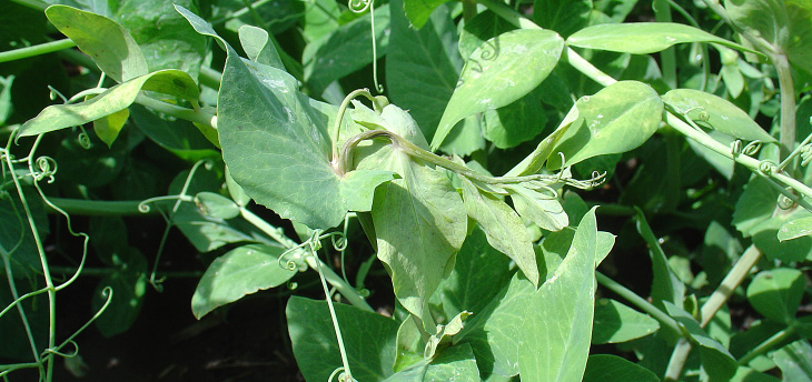
Photo: K. McPhee, NDSU
AUTHORS: Weidong Chen, Lyndon Porter and Kevin McPhee
SYMPTOMS
• Lesions occur on stems, leaves and pods
• Lesions initially are water-soaked but appear bleached and necrotic as they age
• White, puffy fungal growth (white mold) may appear on lesions
• Mouse-dropping-sized black sclerotia may form on and in infected tissue
• Cool and moist conditions
• Lush vegetative growth
• Heavy canopy
IMPORTANT FACTS
• Sclerotia can survive for many years in soil
• Pathogen infects most broadleaf crops
• Plant-to-plant spread can occur by physical contact
• Management tools include clean seed, fungicide applications, rotation to cereal crops and irrigation management
Viruses
Alfalfa mosaic
Alfalfa mosaic virus
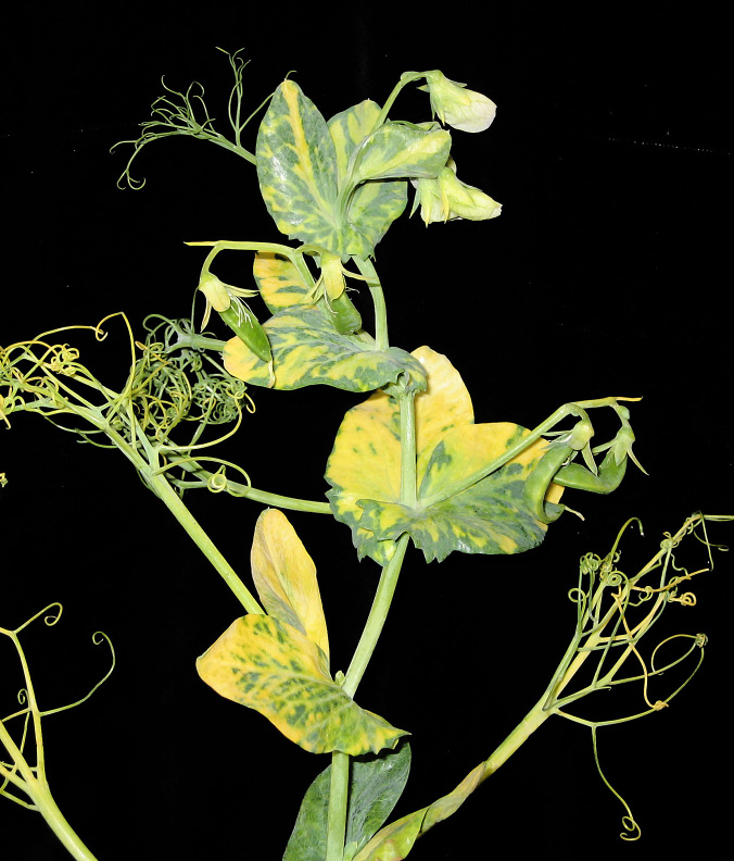
FIGURE 1 – Yellow mottling of foliar tissue
Photo: L. Porter, USDA-ARS Prosser, WA
AUTHORS:Lyndon Porter
SYMPTOMS
• Yellow mottling of foliar tissue (not always prominent)
• Purple or brown streaks in leaf veins
• Dead tissue on leaf or stem
FACTORS FAVORING DEVELOPMENT
• Presence of pea and green peach aphids, which transmit the virus
• Proximity to alfalfa fields
IMPORTANT FACTS
• Pea, green peach, foxglove, bean and potato aphids transmit the virus
• No resistant cultivars are available
• Insecticides may reduce secondary spread of virus by killing vectors (aphids)
• Can be confused with pea streak virus
Bean leaf roll or pea leaf roll
Bean leaf roll virus
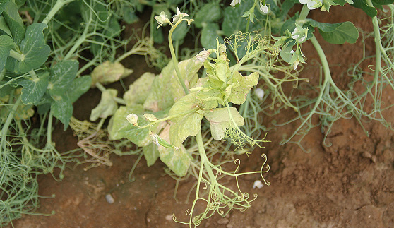
FIGURE 1 – Yellow, distorted and twisted leaves
Photo: L. Porter, USDA-ARS Prosser, WA
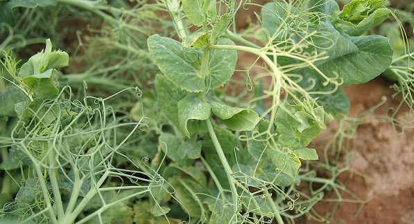
FIGURE 2 – Down-curled leaves
Photo: L. Porter, USDA-ARS Prosser, WA
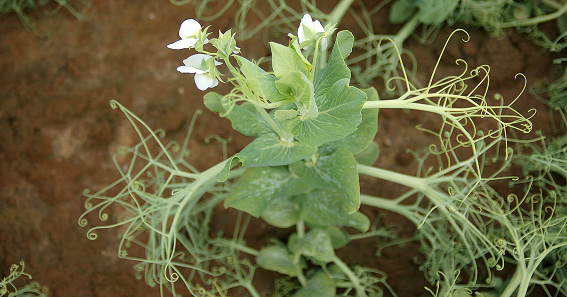
FIGURE 3 – Yellow and distorted new growth; old growth is normal
Photo: L. Porter, USDA-ARS Prosser, WA
AUTHORS: Lyndon Porter
SYMPTOMS
• Plants are yellow and stunted
• New tissue is distorted and twisted while old growth may be normal
• Leaflets curl downward and are brittle
• Presence of pea aphids transmitting the virus
IMPORTANT FACTS
• Virus is not seed-transmitted
• Often occurs with pea enation mosaic virus
• Later infections are less likely to have an impact on yield
• Cultivars with resistance may be available
• Can be confused with other viruses, root rots, herbicide damage or abiotic stress
Pea enation mosaic
Pea enation mosaic virus
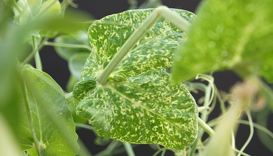
FIGURE 1 – Leaf with mosaic pattern of white/clear spots (windows)
Photo: L. Porter, USDA-ARS Prosser, WA
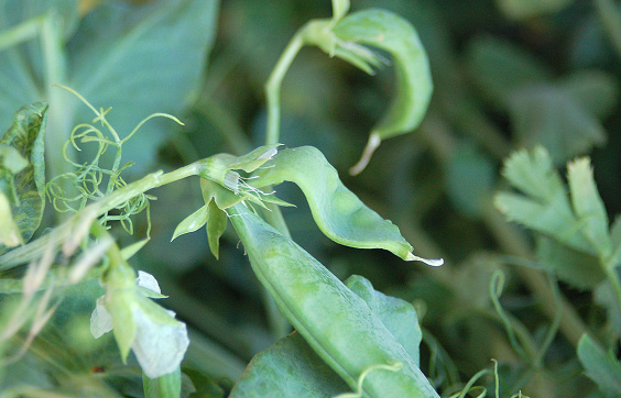
FIGURE 2 – Misshapen pods
Photo: L. Porter, USDA-ARS Prosser, WA
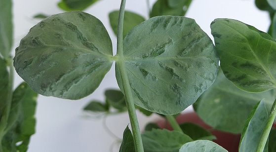
FIGURE 3 – Enations (bumps) on leaf
Photo: L. Porter, USDA-ARS Prosser, WA
SYMPTOMS
• Leaves may be brittle and have a mosaic of green and yellow rough bumps (enations), translucent spots or clear veins
• Pods may be distorted and fill poorly
FACTORS FAVORING DEVELOPMENT
• Presence of pea aphids transmitting the virus
IMPORTANT FACTS
• Virus is not seed-transmitted
• Often occurs with bean leaf roll virus
• Early infections more severely impact yield than late infections
• Insecticides may reduce secondary spread of virus by killing vectors (aphids)
• Can be confused with other viruses, herbicide damage
Pea seedborne mosaic
Pea seedborne mosaic virus

FIGURE 1 – Deformed growth
Photo: L. Porter, USDA-ARS Prosser, WA
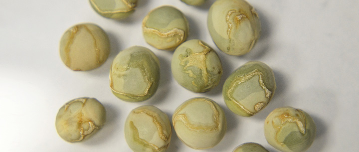
FIGURE 2 – Seed with water soaking and scarring symptoms
Photo: A. Beck, NDSU
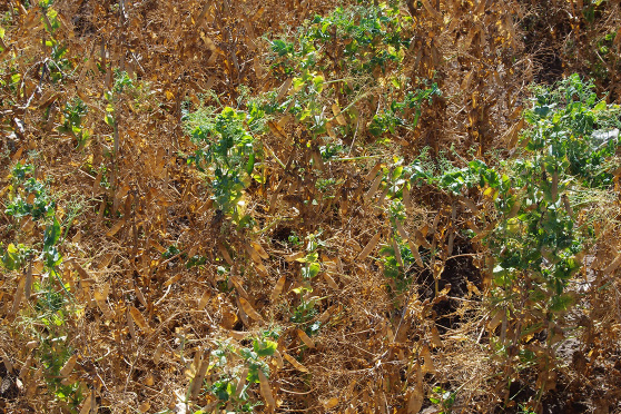
FIGURE 3 – Delayed maturity of infected plants
Photo: M. Wunsch, NDSU
AUTHORS: Lyndon Porter, Kevin McPhee and Julie Pasche
SYMPTOMS
• Leaves may curl downward
• Plants are stunted with a rosette appearance on new growth
• Pods may be deformed and fill poorly
• Seed may be water-soaked, scarred or cracked
• Maturity of infected plants is delayed
• Presence of pea, green peach or potato aphids, which can transmit the virus
• Infected seed
IMPORTANT FACTS
• Virus is readily seed-transmitted
• Virus infects many plants, including lentil, chickpea, alfalfa and vetch
• Manage by planting virus-free seed and resistant cultivars
• Insecticides may reduce secondary spread of virus by killing vectors (aphids)
• Can be confused with other viruses or herbicide damage
Pea streak
Pea streak virus
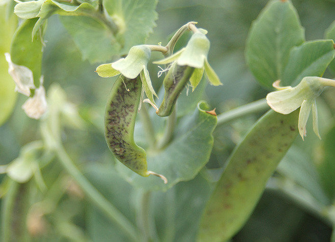
FIGURE 1 – Malformed pea pods with blistering
Photo: L. Porter, USDA-ARS Prosser, WA
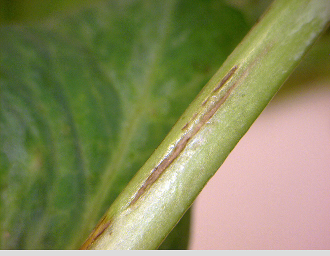
FIGURE 2 – Purple sunken streaks on infected plants
Photo: L. Porter, USDA-ARS Prosser, WA
SYMPTOMS
• Purple to brown streaks on leaves, stems and pods
• Leaf-yellowing and dieback of growing tips
• Pods may appear blistered, deformed and fill poorly
• Streaks on pods differ in size and shape and often are sunken
FACTORS FAVORING DEVELOPMENT
• Presence of pea or green peach aphid transmitting virus
IMPORTANT FACTS
• Virus is not seed-transmitted
• Virus also can infect alfalfa, red and white clover, and vetch
• Rarely associated with significant damage in pea fields
• Insecticides may reduce secondary spread of virus by killing vectors (aphids)
• Can be confused with other viruses, herbicide or abiotic damage
May 2016


