Dry Edible Bean Disease Diagnostic Series (PP1820, Dec. 2016)
Root Diseases
Fusarium root rot
Fusarium solani
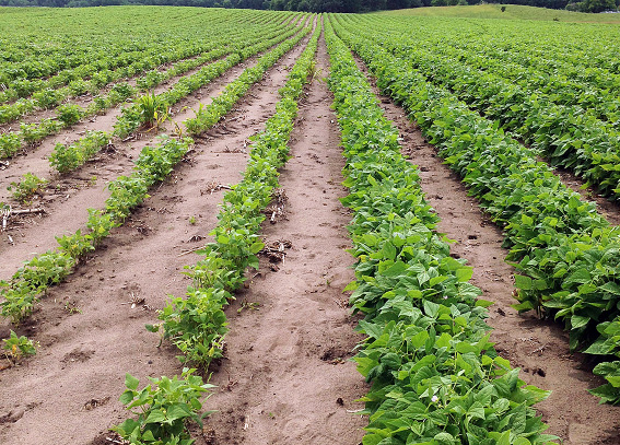
FIGURE 1 – Susceptible (L) and moderately resistant (R) bean varieties under heavy Fusarium root rot pressure
Photo: R. Harveson, Univ. of Nebraska
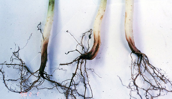
Photo: R. Harveson, Univ. of Nebraska
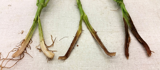
Photo: R. Harveson, Univ. of Nebraska
AUTHORS: Jessica Halvorson, Chryseis Tvedt, Julie Pasche, Bob Harveson and Sam Markell
SYMPTOMS
• Reddish-brown below-ground lesions
• Lesions may extend up the main root and hypocotyl
• Internal brown to red discoloration may be visible
• Yellow and stunted above-ground symptoms
FACTORS FAVORING DEVELOPMENT
• Cool and wet soils after planting
• Compacted soils and plant stress
IMPORTANT FACTS
• Soybeans and other pulse crops may be hosts
• May appear in circular patterns in a field
• Often found in a complex of other root rots
• Fungicide seed treatments may be effective early in the season
• Can be confused with other root rots and abiotic stresses
Pythium diseases
Pythium spp.
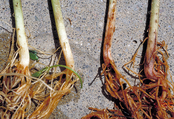
FIGURE 1 – Water-soaking symptoms on roots and hypocotyls (R) and healthy root (L)
Photo: R. Harveson, Univ. of Nebraska
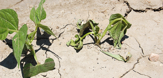
FIGURE 2 – Wilting and death of a young bean plant
Photo: R. Harveson, Univ. of Nebraska
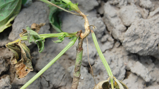
FIGURE 3 – Pythium blight-phase causing necrosis of stems and petioles
Photo: J. Pasche, NDSU
SYMPTOMS
• Initial root rot symptoms appear as elongated, water-soaked necrotic areas on roots or hypocotyls, sometimes extending above soil line
• Wilting and death of plants (damping off)
• Symptoms on above-ground tissues (blight phase) may occur after extended conditions of rain, irrigation, high humidity or high moisture
• High levels of soil moisture
• Disease incidence often is greater where water accumulates in fields
IMPORTANT FACTS
• Cool-weather species (most active below 75 F) include P. ultimum, while warm-weather species (80 to 95 F) include P. myriotylum and P. aphanidermatum
• The pathogens survive in soil for years and can be moved with soil
• Any area of the plant in contact with the soil may become infected, resulting in water-soaked areas of the stem or upper branches (blight-phase)
• Can be confused with other root rots, wilts and white mold (blight-phase only)
Rhizoctonia root rot
Rhizoctonia solani
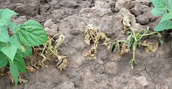
FIGURE 1 – Stunting, wilting and premature death
Photo: G. Yan. NDSU
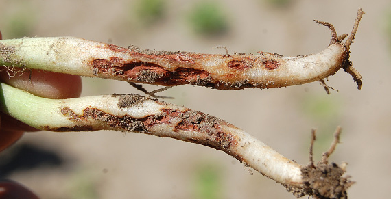
FIGURE 2 – Sunken reddish-brown cankers
Photo: R. Harveson, Univ. of Nebraska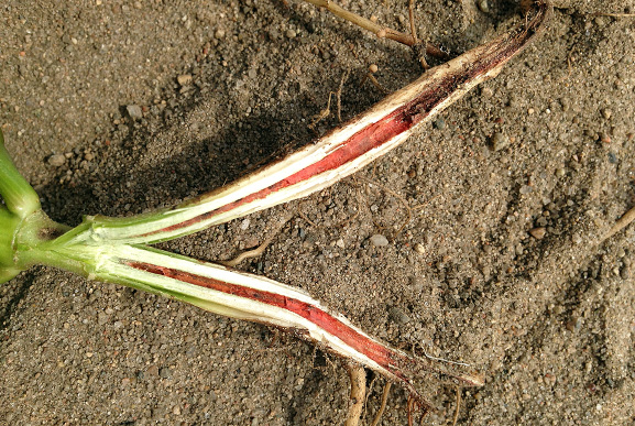
FIGURE 3 – Brick-red discoloration in pith
Photo: J. Pasche, NDSU
AUTHORS: Jessica Halvorson, Julie Pasche, Bob Harveson and Sam Markell
SYMPTOMS
• Stunting and premature death of plants in field
• Lesions or cankers with reddish-brown borders on roots and base of stem
• Internal brick-red discoloration of pith
• Moderate to high soil moisture
• Cool, compacted soil
IMPORTANT FACTS
• Soybeans, sugar beets, potatoes, pulse crops and some weeds are hosts
• Often found in a complex with other root rots
• Fungicide seed treatments may help manage disease early in the growing season
• Can be confused with other root rots and abiotic stresses
Soybean cyst nematode (SCN)
Heterodera glycines
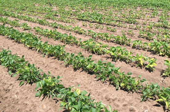
Photo: G. Yan. NDSU
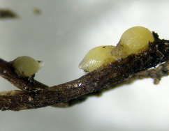
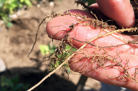
FIGURE 2 – Small cream-colored females on dry bean roots
Photo: G. Yan, NDSU
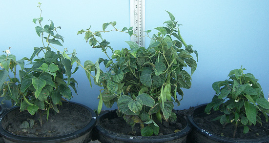
FIGURE 3 – Stunting of pinto bean growing in pots with different levels of SCN; no SCN (C); 5,000 eggs/100cc (L); 10,000 eggs/cc of SCN (R)
Photo: S Poromarto, NDSU
AUTHORS: Julie Pasche, Guiping Yan, Berlin Nelson, Sam Markell and Bob Harveson
SYMPTOMS
• Plants can be infected with no above-ground symptoms
• Stunted or yellow areas of the field
• Small (1/32 to 1/6 inch) cream-colored and lemon-shaped cysts on roots
FACTORS FAVORING DEVELOPMENT
• Rotation with soybeans
• Light soil texture
• High soil pH
• Warm and dry soil
IMPORTANT FACTS
• Soybeans and dry edible beans are hosts
• Dirty equipment, flooding and wind erosion are SCN dispersal mechanisms
• All market classes are hosts
• Research indicates that kidney beans are the market class most susceptible to SCN and black beans are the least susceptible
Soybean cyst nematode soil sampling
Heterodera glycines
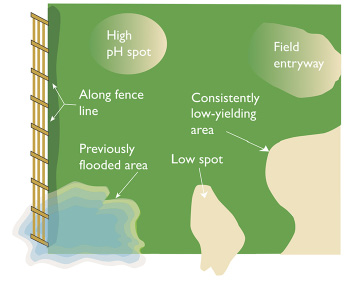
FIGURE 1 – High-risk spots for SCN
Courtesy Iowa State University
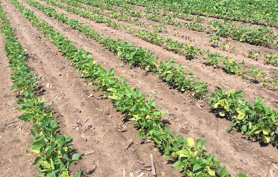
FIGURE 2 – SCN causing yellowing and stunting in kidney beans
Photo: G. Yan, NDSU
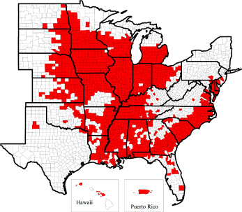
FIGURE 3 – Counties positive for SCN (detected on soybeans) as of 2014
Courtesy Iowa State University
AUTHORS: Sam Markell, Guiping Yan, Berlin Nelson, Julie Pasche and Bob Harveson
WHY SOIL SAMPLE
• SCN is a microscopic worm that lives in the soil and parasitizes roots
• Soil sampling is the most reliable way to detect SCN
WHEN TO SAMPLE
• In late summer/fall (before or after harvest), when SCN population is highest and more easily detected
WHERE TO SAMPLE
• Anything that moves soil can move SCN
• Concentrate sampling in areas where SCN is likely to be introduced or develop, especially field entrances
• Aim for the roots, dig 6 to 8 inches deep, take 10 to 20 samples, mix and send to a lab
WHAT RESULTS MEAN
• Results are presented as eggs/100 cc, which is the number of nematode eggs in approximately 3.4 ounces of soil
• Low levels (for example, 50 or 100 eggs/100 cc) could be false positives and should be viewed with caution
Stem and Wilt Diseases
Bacterial wilt
Curtobacterium flaccumfaciens pv. flaccumfaciens
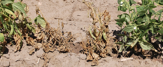
Photo: R. Harveson, Univ. of Nebraska
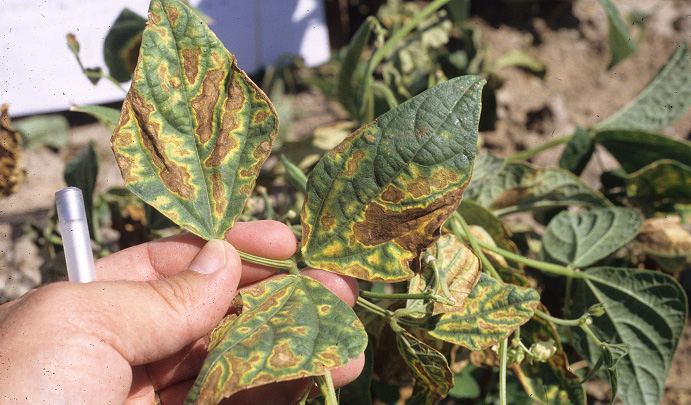
Photo: R. Harveson, Univ. of Nebraska
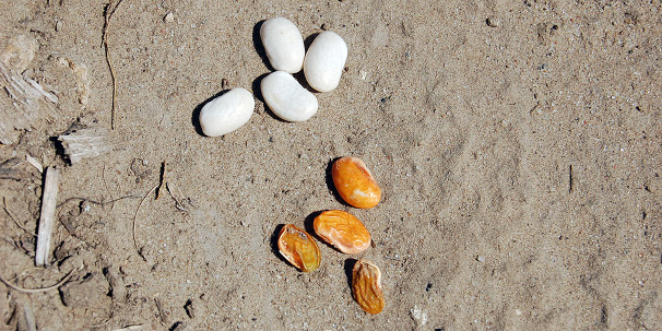
FIGURE 3 – Shriveled, orange-stained seeds (bottom) and healthy seeds (top) obtained from the same infected plant
Photo: R. Harveson, Univ. of Nebraska
AUTHORS: Bob Harveson, Sam Markell and Julie Pasche
SYMPTOMS
• Leaf wilting during periods of warm, dry weather or periods of moisture stress
• Interveinal, necrotic lesions which may be surrounded by bright yellow borders
• Seeds from surviving infected plants often will shrivel and be stained yellow or orange
• Very hot air temperatures (greater than 90 F), with wet or humid conditions
IMPORTANT FACTS
• Wilt pathogen survives in bean residue or seeds from previous year
• Infected seeds are primary mechanism of long-distance movement
• Wet weather, hail, violent rain and windstorms help the pathogen spread within and between fields
• Can be confused with root rots and other bacterial pathogens; foliar symptoms of bacterial wilt tend to be more wavy or irregular than common bacterial blight lesions and do not include water-soaking
Fusarium yellows (wilt)
Fusarium oxysporum f. sp. phaseoli
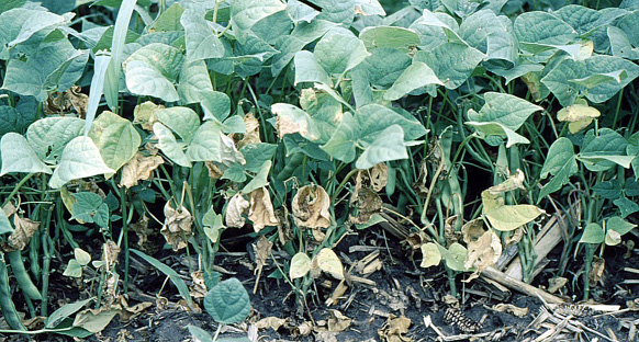
FIGURE 1 – Yellowing and wilting of leaves
Photo: R. Harveson, Univ. of Nebraska
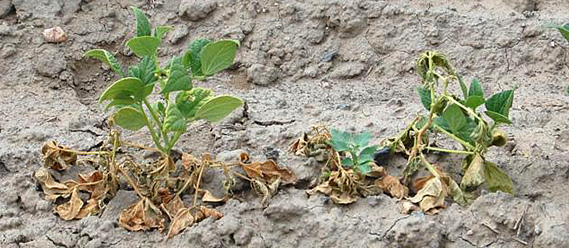
FIGURE 2 – Permanent wilting and death of severely affected plants
Photo: R. Harveson, Univ. of Nebraska
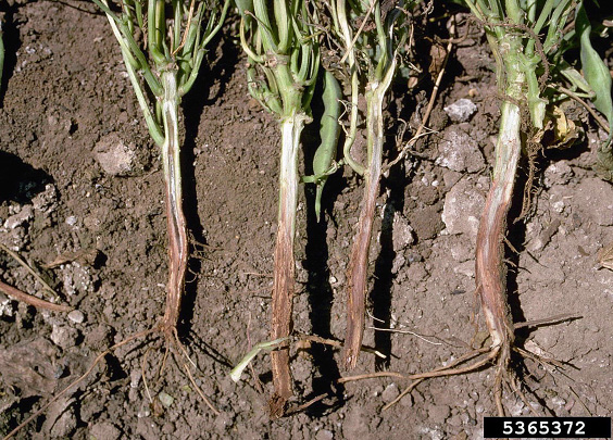
FIGURE 3 – Vascular discoloration of plants affected by Fusarium wilt
Photo: R. Harveson, Univ. of Nebraska
SYMPTOMS
• Foliar symptoms first appear as yellowing and wilting of older leaves, followed by younger leaves if the disease progresses
• Severely affected plants may wilt permanently
• Vascular discoloration of roots and hypocotyl tissues is primary diagnostic symptom; degree of discoloration varies in intensity depending on cultivar and environmental conditions
FACTORS FAVORING DEVELOPMENT
• High temperature stress (greater than 86 F)
• Dry soil conditions
• Soil compaction
IMPORTANT FACTS
• Fusarium wilt often causes more dramatic symptoms than Fusarium root rot infections
• Unlike Fusarium root rot infections, Fusarium wilt seldom kills plants
• Death with wilt can occur before or after pod set
• Fusarium wilt can induce maturity two to three weeks earlier than normal
• Can be confused with other root rot and wilt diseases
Stem rot
Unknown sterile white basidiomycete (SWB)
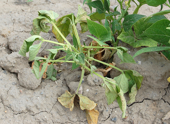
FIGURE 1 – Wilting symptoms characteristic of SWB infection
Photo: R. Harveson, Univ. of Nebraska
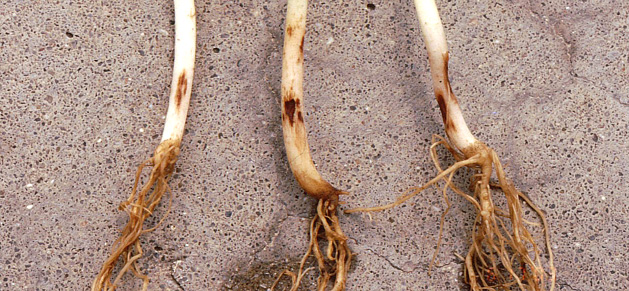
FIGURE 2 – Small light brown lesions (L), moderate lesions (C) and large dark brown to black sunken lesions (R)
Photo: R. Harveson, Univ. of Nebraska
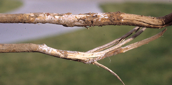
FIGURE 3 – White mycelial strands of SWB and soil adhering to stems of infected plants
Photo: R. Harveson, Univ. of Nebraska
AUTHORS: Bob Harveson, Sam Markell and Julie Pasche
SYMPTOMS
• Wilting and death of young plants first observed after emergence
• On less severely affected plants, small lesions may be on hypocotyls
• Severe infection also can include sunken gray to black cankers on hypocotyls and stems
• White mycelial strands may grow over lesions or into stem piths; soil will adhere to stems when wilted plants are removed
FACTORS FAVORING DEVELOPMENT
• High soil temperatures, but has been reported to cause disease from 60 to 95 F
IMPORTANT FACTS
• Thought to have many hosts
• Can survive at least one year in soils, likely in colonized residue of weeds or other susceptible crops
• Can be confused with other root rots, wilts and white mold
White mold
Sclerotinia sclerotiorum
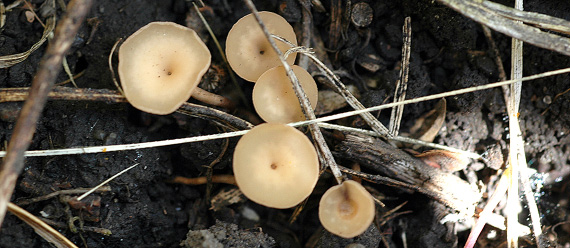
FIGURE 1 – Small tan mushrooms (apothecia) about ¼ inch in diameter emerge from hard, black structures (sclerotia)
Photo: S. Markell, NDSU
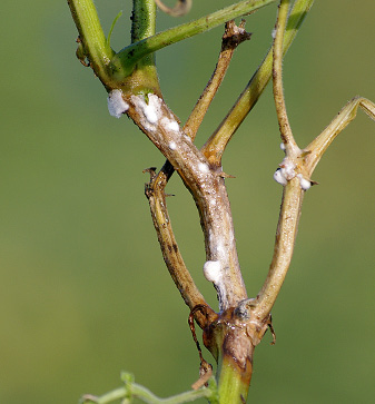
FIGURE 2 – Enlarging tan lesions with white fungal growth
Photo: M. Wunsch, NDSU
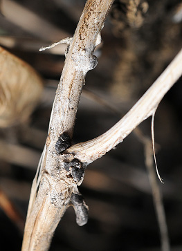
FIGURE 3 – Mature stem lesion with dried-bone appearance, white fungal growth and black sclerotia
Photo: S. Markell, NDSU
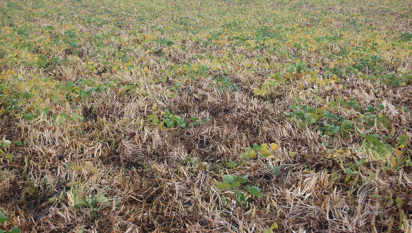
FIGURE 4 – Severe white mold damage
Photo: R. Harveson, Univ. of Nebraska
AUTHORS: Julie Pasche, Bob Harveson and Sam Markell
SYMPTOMS
• Water-soaked lesion that becomes tan as it enlarges
• Stem lesions will dry out, lighten in color and tissue may shred
• White fungal growth and hard black sclerotia may form in or on stem
FACTORS FAVORING DEVELOPMENT
• Wet soils prior to bloom; allows sclerotia to germinate and release spores
• Cool daytime temperatures (60 to 70F) during and after bloom
• Long periods of canopy wetness and/or frequent rainfall during bloom
• Lush plant growth
IMPORTANT FACTS
• All broadleaf crops and many weeds are susceptible to white mold
• Plants are only susceptible when in bloom
• Preventative fungicide applications may be economically viable
• Can be confused with wilt diseases or abiotic stress
Foliar Diseases
Anthracnose
Colletotrichum lindemuthianum
Photos: S. Markell, NDSU
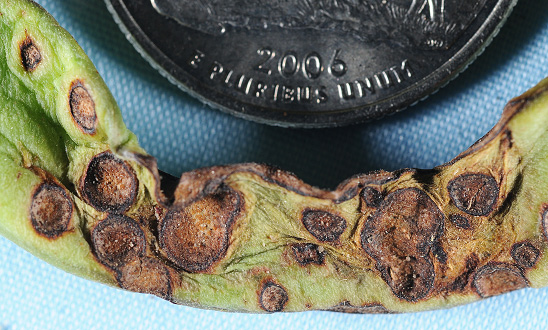
Photo: S. Markell, NDSU
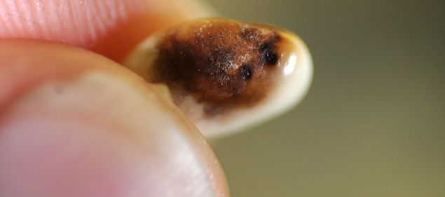
Photo: S. Markell, NDSU
AUTHORS: Jessica Halvorson, Sam Markell, Julie Pasche and Bob Harveson
SYMPTOMS
• Can occur on all above-ground plant parts
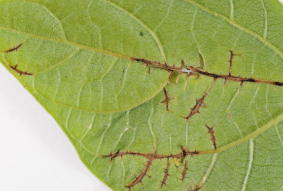
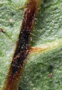
• Leaf vein and petiole lesions are dark and slender
• Pod lesions begin as small brown spots, enlarge to become circular and sunken
• Infected seeds may appear discolored and have necrotic lesions
• White fungal growth or cream-salmon-colored spore masses may be visible in lesions
FACTORS FAVORING DEVELOPMENT
• Infected seed
• Cool (55 to 80 F) temperatures
• Frequent rain or thunderstorms
IMPORTANT FACTS
• Pathogen is seed-borne and wind-dispersed
• Spread can occur by splashing water
• Pathogen can spread by animals, people or machinery moving through fields when foliage is wet
• Planting certified disease-free seed is best way to prevent the disease
• Can be confused with bacterial blights
Bacterial brown spot
Pseudomonas syringae pv. syringae
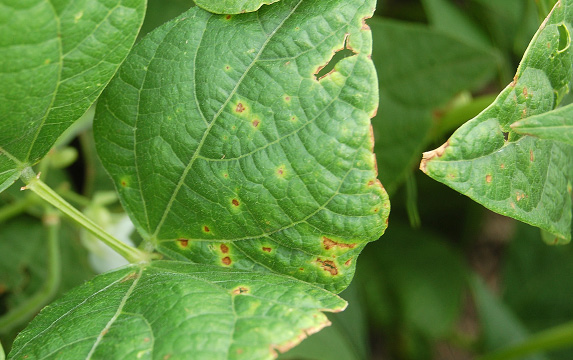
Photo: R. Harveson, Univ. of Nebraska
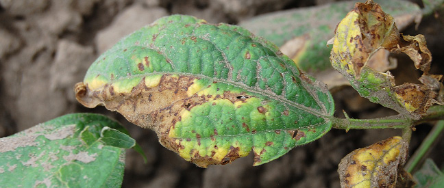
Photo: R. Harveson, Univ. of Nebraska
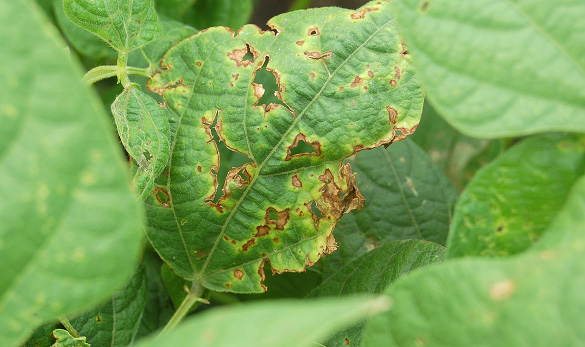
FIGURE 3 – Older lesions with holes after necrotic tissues fell out
Photo: R. Harveson, Univ. of Nebraska
SYMPTOMS
• Small, circular, brown lesions, often surrounded by a narrow yellow zone (not always present)
• Lesions may coalesce to form linear necrotic streaks between leaf veins
• Centers of old lesions dry and fall out, leaving tattered strips or “shot holes”
• May infect leaves, pods and seeds
• Warm air temperatures (80 to 85 F) with wet or humid conditions
• Storms that damage plants (hail, high wind)
• Planting infected seeds favors early infection and disease spread
IMPORTANT FACTS
• Pathogen survives in seed, residue and on other living hosts
• Wet weather, hail, violent rain and windstorms spread the pathogen
• Can be confused with other bacterial blights: necrotic area is similar in size to halo blight but smaller than common bacterial blight; yellow margin (halo) is narrow and bright as with common blight, but halo blight’s is larger, faint
Bean common mosaic
Bean common mosaic virus (BCMV)
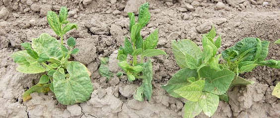
FIGURE 1 – Mosaic, blistering and distortion (elongation) of leaves of affected plants
Photo: R. Harveson, Univ. of Nebraska
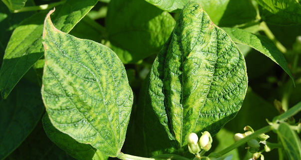
FIGURE 2 – Vein banding of leaves on an infected plant
Photo: R. Harveson, Univ. of Nebraska
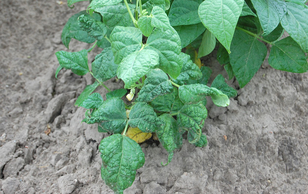
Photo: R. Harveson, Univ. of Nebraska
AUTHORS: Bob Harveson, Julie Pasche and Sam Markell
SYMPTOMS
• Light and dark green mosaics and/or leaf malformation
• Downward rolling or cupping of leaves
• Vein banding, and stunting, necrosis or premature death
FACTORS FAVORING DEVELOPMENT
• Disease development dependent on susceptibility of cultivars and presence of aphids as vectors
• Yield losses more severe after early infections
IMPORTANT FACTS
• Type and severity of symptoms depend on host cultivar, virus strain and environment
• BCMV is spread among production areas by planting infected seed
• Several aphid species transmit BCMV
• More than 10 strains of BCMV are known
• Can be confused with other viruses, herbicide damage or plant stress
Common bean rust
Uromyces appendiculatus
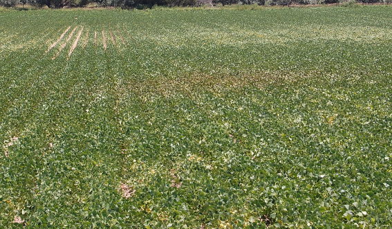
Photo: R. Harveson, Univ. of Nebraska
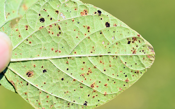
FIGURE 2 – Cinnamon-brown (uredinia) and black (telia) rust pustules
Photo: S. Markell, NDSU
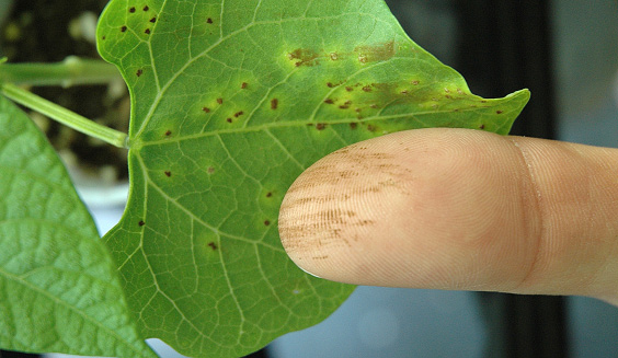
FIGURE 3 – Dusty cinnamon-brown spores rubbed off pustule with yellow halo
Photo: S. Markell, NDSU
AUTHORS: Sam Markell, Bob Harveson and Julie Pasche
SYMPTOMS
• Small (1/16 inch) cinnamon-brown pustules that may have a yellow halo
• Pustules turn black at end of growing season
• Usually first observed in areas of a field with concentrated infection, called “hot spots”
• Close proximity to a field that had rust the previous year
• Frequent heavy dews
• Moderate to warm temperatures (65 to 85 F)
• Factors favoring wet microclimates: lush plant growth, close to shelter belts, etc.
IMPORTANT FACTS
• Pathogen is specific to edible beans
• Infection may occur at any time and spread very quickly
• Fungicides applied after detection may be economically viable
• Pathogen has different races, which may overcome resistance
• Can be confused with soil splash, brown spot and halo blight
Common bacterial blight
Xanthomonas campestris pv. phaseoli
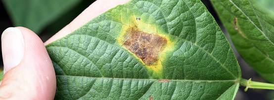
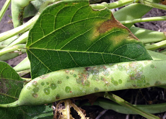
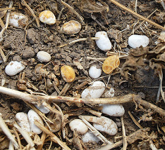
FIGURE 1 – Large necrotic lesions with narrow yellow borders
Photo: S. Markell, NDSU
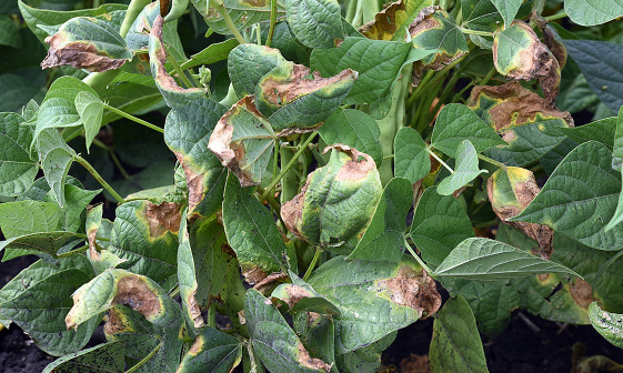
FIGURE 2 – Severely damaged leaves appearing burned or scorched
Photo: S. Markell, NDSU
AUTHORS: Bob Harveson, Julie Pasche and Sam Markell
SYMPTOMS
• Leaves, pods and seeds can be infected
• Initial symptoms: small water-soaked spots on the underside of leaves
• Spots enlarge and coalesce to form large necrotic areas with a narrow, bright yellow border
• Severely damaged leaves appear burned and remain attached at maturity
FACTORS FAVORING DEVELOPMENT
• Warm air temperatures (80 to 90 F) with wet or humid conditions
• Storms that damage plants (hail, high wind)
• Planting infected seeds favors early infection and disease spread
IMPORTANT FACTS
• Bacteria survive in fields on infected seed or bean tissues
• Pathogen can spread by animals, people or machinery moving through fields when foliage is wet
• Can be confused with anthracnose (pod infection) and bacterial diseases; yellow margin (halo) is similar in color and brightness to bacterial brown spot but necrotic area is larger
Halo blight
Pseudomonas syringae pv. phaseolicola
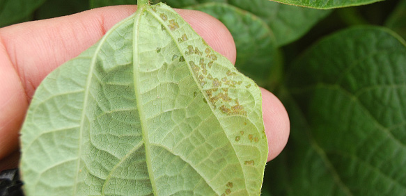
FIGURE 1 – Small water-soaked spots on underside of leaf
Photo: R. Harveson, Univ. of Nebraska
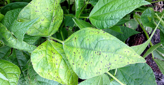
FIGURE 2 – Broad yellow-green halo surrounding small necrotic spot
Photo: J. Pasche, NDSU
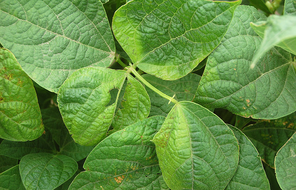
FIGURE 3 – Severe infection and the beginning of a systemic chlorosis in plants
Photo: R. Harveson, Univ. of Nebraska
AUTHORS: Bob Harveson, Julie Pasche and Sam Markell
SYMPTOMS
• Begins with small water-soaked spots that become necrotic
• Broad yellow-green halo may develop around necrotic spots
• In severe cases, a general systemic chlorosis may develop in infected plants
• Also may infect pods and seeds
FACTORS FAVORING DEVELOPMENT
• Cool air temperatures (68 to 72 F) with wet or humid conditions
• Planting infected seeds favors early infection and disease spread
• Storms with high winds, rain or hail will damage plants and spread pathogen from plant to plant
IMPORTANT FACTS
• Yellow-green chlorotic halo more pronounced at cool temperatures, less noticeable above 75 F
• Pathogen can spread by animals, people or machinery moving through fields when foliage is wet
• Can be confused with other bacterial blights; necrotic area is similar in size to bacterial brown spot but halo is much larger and a fainter yellow-green


December 2016

